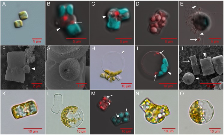
Fig 3.
The life cycle stages of T. pseudonana (A-K), T. weissflogii (L) and C. cryptica (M-O) imaged using scanning electron microscopy (SEM), light (LM), and confocal microscopy (CFM). A: Two vegetative cells (LM, CCMP1335). B: Oogonium displaying separation of thecae (arrowhead) and putative pycnotic nucleus indicated by the arrow (CFM, CCMP1015). C: Oogonium sharply bending at the thecae junction. Arrowhead indicates protrusion of the plasma membrane (CFM, CCMP1335). Oogonia images are representative of 38 total images. D: Spermatogonium containing multiple spermatocytes seen as individual red (DNA stained) clusters (CFM, CCMP1335); representative of 8 images. E: Motile spermatocytes (in red, arrow) with moving flagella (arrowheads, CFM, CCMP1335, representative of 10 images). F-G: SEM images of spermatocytes (arrowhead) attached to early oogonia (SEM, CCMP1335, representative of 20 images). H,I: Auxospores; representative of 60 images in CCMP1015 (H, LM) and CCMP1335 (I, CFM) showing bulging where mother valve was attached (arrowhead). Two nuclei are visible in red following non-cytokinetic mitosis. J: Small parental cell (arrow) with initial cells produced by sexual reproduction to the left (partial valve view) and right (girdle view) indicated by the arrowheads (SEM, CCMP1335). K: 7 x 12 μm initial cell (LM); j and k representative of 12 images of CCMP1335. L: T. weissflogii auxospore (LM); representative of 12 similar images. M: C. cryptica spermatogonium (upper left) and vegetative cell (lower right). CFM shows stained DNA (red, arrow) and multiple nuclei in the spermatogonium. Arrowheads indicate chlorophyll autofluorescence (green). Oogonium (N, representative of 6 images) and auxospore (O, representative of 4 similar images) of C. cryptica (LM). Confocal microscopy images (b-e, i, m) show chlorophyll autofluorescence (green) and Hoescht 33342 stained DNA (red). Scale bars: A: 5 μm; B-E: 5 μm; F: 2 μm; G: 1 μm; H-O: 10 μm.

