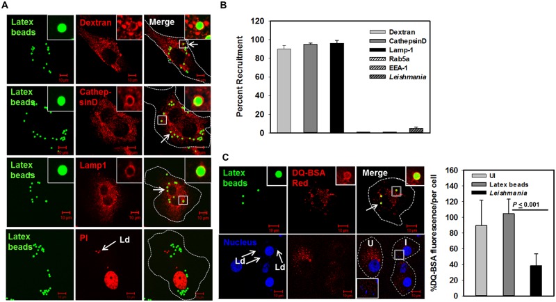
Fig 6. Leishmania donovani inhibits its transport to lysosomes in macrophages.
A. Lysosomes of THP-1 differentiated human macrophages were prelabeled with internalization of Dextran Texas Red for 24 h and subsequently, cells were infected with Fluoresbrite-YG-latex beads (Green) as described in Materials and Methods (upper panel). Similarly, lysosomes of THP-1 differentiated human macrophages were labeled with Green fluorescent labeled latex beads and cells were immune-stained with anti-CathepsinD (middle panel) or anti-Lamp1 (middle panel) antibody as described in Materials and Methods. THP-1 differentiated human macrophages were coinfected with Leishmania and Green fluorescent labeled latex beads and chased for 24 h at 37°C and their localization was determined by confocal microscopy (lower panel). Leishmania and macrophage nucleus were stained with propidium iodide (Red). All results are representative of three independent experiments. B. Results are represented as mean ± S.D. of three independent experiments and expressed as percentage of latex-bead containing phagosomes positive for indicated markers after counting 100 cells. C. THP-1 differentiated human macrophages were infected with Leishmania or Green fluorescent labeled latex beads. Cells were incubated for 24 h at 37°C and lysosomes were labeled with DQ-BSA Red as described in Materials and Methods. Leishmania and macrophage nucleus were stained with Hoechst (Blue). Cells were viewed in an LSM 510 Meta confocal microscope using an oil immersion objective. Results are expressed as mean percentage of total fluorescence per cell ± S.D. of 100 independent indicated cells.

