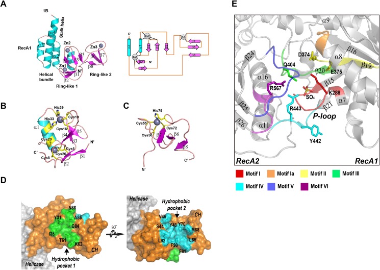
Fig 3. Key structural features of MERS-CoV nsp13.
(A) left, ribbon model of the CH and Stalk domains of MERS-CoV nsp13 colored by secondary structural elements; right, 2-D topology graph of the CH domain. (B) ribbon model of N-terminal Ring module, and (C) C-terminal Ring module of the CH domain. His/Cys residues participating in zinc coordination are highlighted in yellow. (D) Surface representation of CH domain (orange) of MERS-CoV nsp13, two hydrophobic pockets equivalent to protein interaction interfaces on Upf1 are highlighted in green (pocket 1) and cyan (pocket 2). (E) The ATPase active site between RecA1 and RecA2 domains. The conserved helicase motifs are highlighted with different colors.

