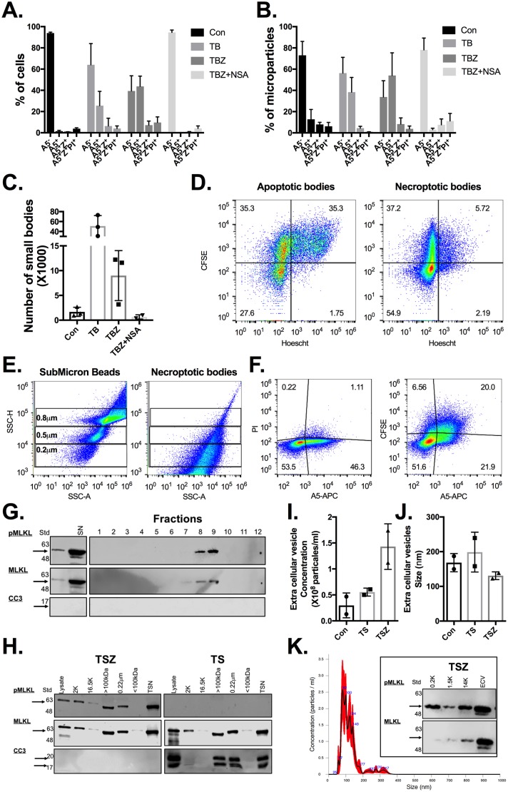
Fig 4. Necroptotic cells release PS-exposed ECVs and pMLKL.
(A-C) CFSE labeled U937 cells were stimulated for either (i) apoptosis (TB), necroptosis (TBZ), or (ii) left untreated (Con). Three hours after stimulation, cells (A) and microparticles (B) were divided by their A5, Z, and PI staining, and the geometric mean of CFSE fluorescence in the different populations was then calculated using FloJo software. (C) Calculated total number of microparticles in the different treatments. Data are taken from 3 independent experiments. (D) CFSE and Hoescht prelabeled U937 cells were stimulated as above. Three hours after stimulation, apoptotic/necroptotic bodies were isolated and then analyzed for CFSE and Hoescht staining using flow cytometry. (E–F) ECVs from CFSE labeled U937 necroptotic cells were isolated using a size exclusion column (qEV, ZION), (fractions 7–9). (E) ECV (right panel) size was compered to submicron beads of known size (left panel) and (F) further stained for A5 and PI, and then analyzed using flow cytometry. (G) The different isolated fractions (qEV, ZION) from U937 necroptotic cells were concentrated, and the cell death key factors pMLKL and CC3 were detected using western blot. (H) U937 cells were stimulated for apoptosis (TS) or necroptosis (TSZ). Treated cells and supernatants were fractionated as illustrated in S5C Fig. Cell death key factors pMLKL and CC3 were detected in the different fractions using western blot. (I–K) 5 x 10⁶ U937 cells were stimulated for either (i) apoptosis (TB), necroptosis (TBZ), or (ii) left untreated (Con). ECVs from treated supernatants were isolated using ultracentrifuge and their concentration (I) and size (J) were analyzed using NanoSight. (K) Example NanoSight histogram and detection of pMLKL in ECVs. All raw data for the data summarized under this Fig can be found in S4 Data. APC, allophycocyanin; A5, annexin V; CC3, cleaved caspase 3; Con, control; CFSE, carboxyfluorescein succinimidyl ester; ECV, extracellular vesicle; MFI, geometric mean fluorescence intensity; MLKL; mixed lineage kinase domain-like; NSA, necrosulfonamide; PI, propidium iodide; pMLKL, phosphorylated mixed lineage kinase-like; PS, phosphatidylserine; Std, protein ladder; SN, supernatant; SSC, side scatter; TB, TNFα + birinapant; TBZ, TNFα + birinapant + zVAD; TS, TNFα + SMAC mimetic; TSN, total supernatant; TSZ, TNFα + SMAC mimetic + zVAD; Z, Zombie; zVAD, Z-VAD-FMK.

