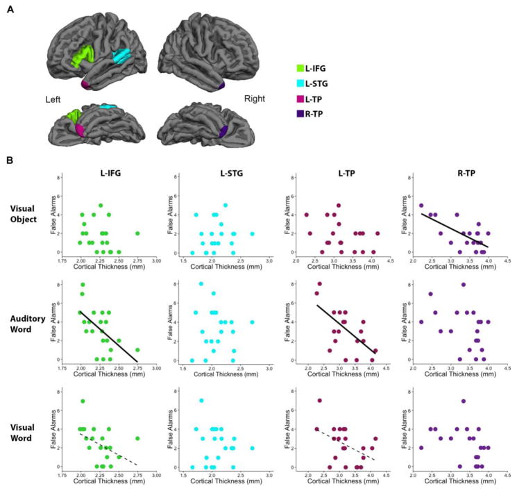
Figure 3. Relationship between mean cortical thickness and false alarms.
(A) Four a priori ROIs are presented in the lateral and inferior view of the left and right hemisphere. (B) Within the PPA cohort (n=21), mean cortical thickness was correlated with false alarms for each stimulus modality. Notably, L-IFG thickness and L-TP thickness correlated with false recognition memory of auditory words and R-TP thickness correlated with false recognition memory of visual objects. Solid lines represent significant correlations (p<0.0125), while dashed lines represent trends (p<0.05). Abbreviations: ROI = Region of Interest, L-IFG = Left Inferior Frontal Gyrus, L-STG = Left Superior Temporal Gyrus, L-TP = Left Temporal Pole, R-TP = Right Temporal Pole.

