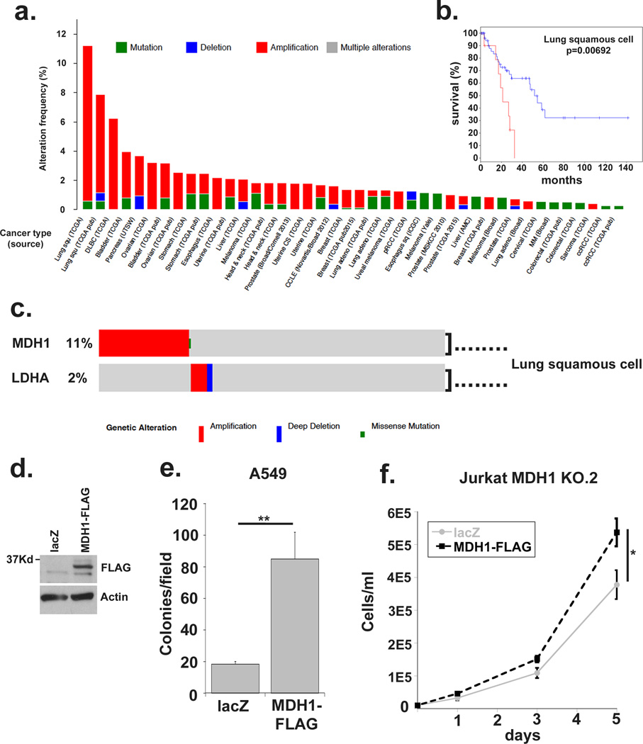
Figure 4. MDH1 is amplified in human cancers and correlates with poor prognosis.
(a) The Cancer Genome Atlas online database was queried using cBioportal software to query MDH1 expression aberrations, including amplification, deletions and mutations in human tumors. (b) The lung squamous cell carcinoma subset was further interrogated to correlate MDH1 amplification with disease free survival. (c) MDH1 and LDHA queried together for genomic aberrations in the lung squamous cell carcinoma dataset. (d) Western blot demonstrating expression of MDH1-FLAG in stably transfected A549 cell lines. (e) Colony counts after 2-week soft agar colony formation assays. The average number of colonies from ten low-magnification microscopic fields from triplicate wells is shown. (f) MDH1 KO.2 Jurkat cells were stably transfected with MDH1-FLAG or lacZ and plated at 10,000 cells per ml. Cell concentration was plotted on days 1, 3 and 5. Data represented are the average and standard deviation of triplicate samples from a representative experiment repeated at least twice. Significance was calculated using the student’s T test (* p<0.01, ** p<0.001).

