
Figure 2.
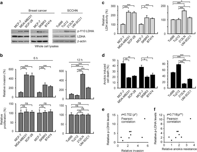
LDHA is phosphorylated at Y10 and activated in a group of highly invasive and anoikis resistant human cancer cell lines. (a) Expression and Y10 phosphorylation levels of LDHA in breast cancer cells (MCF-7, MDA-MB231, SCP-28, SKBR3, BT474) and SCCHN cells (Tu686, Tu212, 212LN, UM-SCC1) shown by western blot analyses. (b) In vitro matrigel invasion assay demonstrates different invasive ability at 6 or 12 h. Proliferation rates at the corresponding time points are shown at the bottom. (c) LDH activity was determined by measuring the oxidation of NADH. (d) Anoikis assay was performed by detecting detachment-induced apoptotic cells using annexin V staining. Cells were cultured on 1% agar-treated dishes for 48 h and detachment-induced cell death was determined by FITC- or PE-annexin V staining. Data are mean ± s.d. from three technical replicates of each sample. P values were determined using two-tailed Student’s t-test for (b–d). (e–f) Pearson correlation between LDHA Y10 phosphorylation and invasive potential (e) or anoikis resistance (f) in the breast cancer and SCCHN cell lines (ns: not significant, *0.01 < P < 0.05, **0.001 < P < 0.01, ***P < 0.001).