
Figure 5.
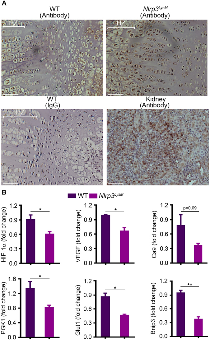
Activation of NLRP3 in myeloid cells impairs chondrocyte responses to hypoxia. (A) Femoral sections from 2-week-old WT or Nlrp3 LysM male mice injected with hydroxyprobe were stained with IgG or hydroxyprobe antibody. A specimen from renal medulla was used as a positive control. Staining is indicated by the brown color. HZ, hypertrophic zone. (B) qPCR analysis of mRNA isolated from mouse epiphyses. Quantitative data were obtained from 2–3 mice/genotype and expressed as the mean ± SEM. *P < 0.05; **P < 0.005.