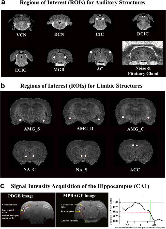
Figure 5.

Representative ROI placements. (a) To measure manganese uptake in the auditory system, ROIs were placed in the left and right dorsal cochlear nuclei (DCNs), ventral cochlear nuclei (VCNs), central nuclei of the inferior colliculus (CICs), external cortices of the inferior colliculus (ECICs), dorsal cortices of the inferior colliculus (DCIC), medial geniculate bodies (MGBs), and auditory cortices (ACs). ROIs were also placed in the left and right anterior pituitary glands and nearby noise. (b) To measure the limbic system, ROIs were placed in the left and right centromedial amygdala (AMGC), the superficial/cortical-like amygdala (AMGS), deep/basolateral amygdala complex (AMGD), nucleus accumbens core (NAC) and shell (NAS), and anterior cingulate cortices (ACCs). (c) To measure the hippocampus, four rectangular bands (white color) were first placed onto the PDGE image. They were then copied onto the corresponding MPRAGE image.
