Figure 4.

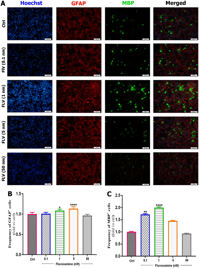
Effect of fluvoxamine on eNSC differentiation to glial and oligodendrocyte cells. (A) Representative immunofluorescence images of differentiated embryonic NSCs stained with GFAP (Glial Fibrillary Acidic Protein, an astrocyte marker, red) or MBP (Myelin Basic Protein, an oligodendrocyte marker, green) and Hoechst (a nuclei marker, blue) in control, and treated groups at day 6. White scale bars indicate 100 𝜇m. (B,C) Quantification of the frequency of GFAP and MBP positive cells in treated groups, normalized to the control group. Results showed that fluvoxamine at 1 and 5 nM concentrations are optimal for differentiation to MBP and GFAP positive cell, respectively. Each group included 3 replicates (n = 3). Values are presented as mean ± SEM for 15 field/well. Statistical analyses were performed by one-way analysis of variance followed by Tukey test. *p < 0.05; **p < 0.01; ***p < 0.001 and ****p < 0.0001.
