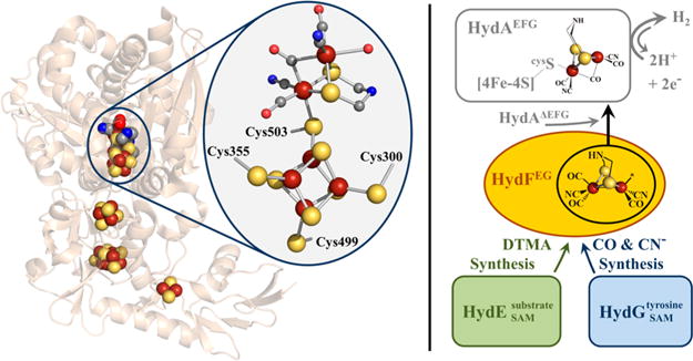
Figure 1.

Left panel. X-ray crystal structure of [FeFe]-hydrogenase from Clostridium pasteurianum I (CpI) (PDB: 3C8Y). The H-cluster is highlighted within the oval. The H-cluster and accessory FeS clusters are depicted as spheres. Color scheme is as follows: iron, rust; sulfur, yellow; carbon, gray; oxygen, red; nitrogen, blue. Right panel. Hypothetical maturation scheme for 2Fe subcluster biosynthesis (see main text for additional details).
