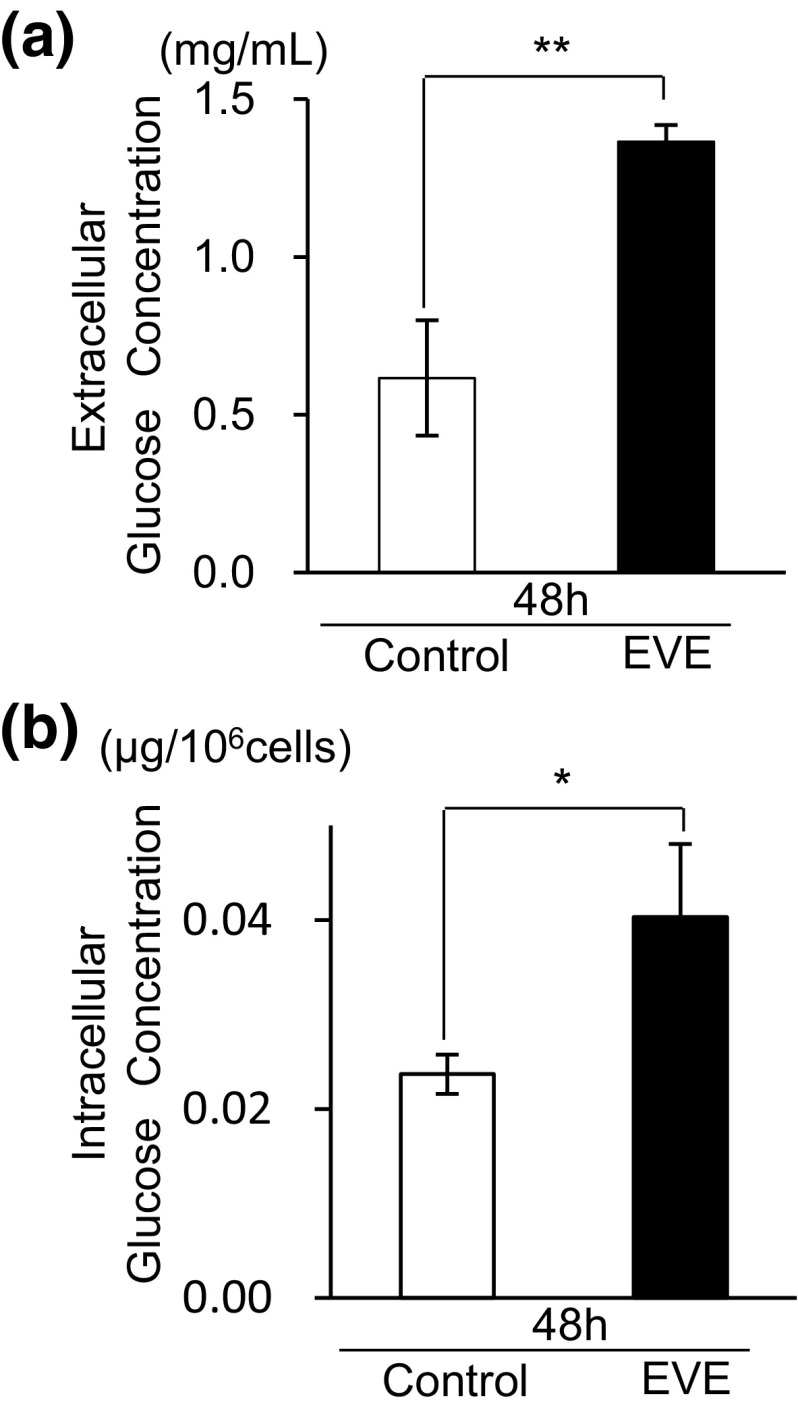
Fig. 1.
Changes in extracellular and intracellular glucose concentrations. At 0 h, 500 ng/mL of everolimus was added to the cultured media (EVE). a The cultured media were collected for measurement of extracellular glucose at 48 h. The original (0 h) concentration of cultured media was 4.5 mg/mL. The open bar represents the glucose concentration of the 48 h control sample and the filled bar denotes a sample exposed to everolimus for 48 h. Data represent the mean ± S.D. (n = 3). Statistically significant difference between EVE and control: **p < 0.01 (Student’s t-test). b Intracellular glucose concentration at 48 h. Open bars represent 48 h control cells and filled bars denote 48 h everolimus-exposed cells. Data represent the mean ± S.D. (n = 3). Statistically significant difference between EVE and control: *p < 0.05 (Student’s t-test)

