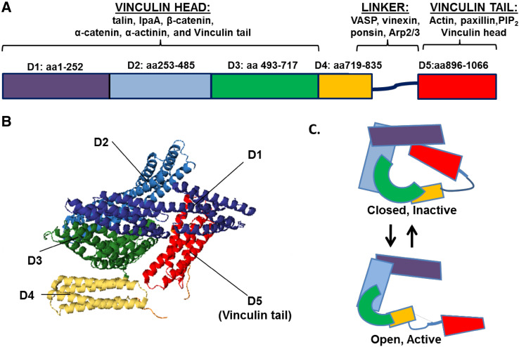
Fig. 1.
Vinculin structure and binding partners. Vinculin is comprised of anti-parallel α-helical bundles organized into five distinct domains. a Domains 1–3 (D1–D3) make up the vinculin head, while domain 5 (D5) encompasses the tail. The binding sites for many proteins interacting with vinculin have been mapped. b The ribbon diagram derived from the human full-length vinculin crystal structure shows vinculin resides in an inactive, closed conformation largely due to tight interactions between D1 and D5. The structure was derived from the PDB coordinates that were supplied by [72]. c A schematic of vinculin in closed, inactive conformation and open, active conformation

