Fig. 1.
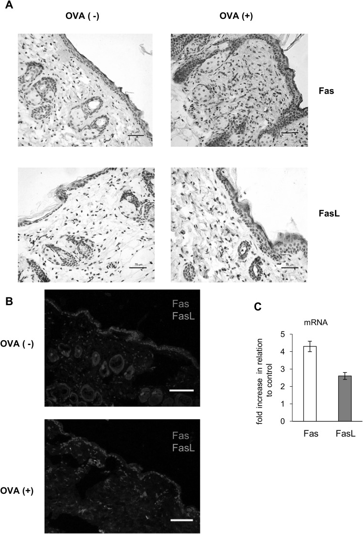
Epicutaneous sensitization with ovalbumin (OVA) induces Fas and FasL expression. a Representative images of Fas and FasL expression identified by immunohistochemistry method in the in paraffin-embedded slides prepared from the skin of C57BL/6 mice, sensitized with saline (OVA−) or ovalbumin (OVA+). The nuclei were counterstained with Harris hematoxylin (violet). Magnification ×200. b Double staining for Fas (red) and FasL (green) by immunohistofluorescence method in the in paraffin-embedded slides prepared from the skin of C57BL/6 mice, sensitized with saline (OVA−) or ovalbumin (OVA+). The nuclei were counterstained with Hoechst 33342 (blue). White scale bar shows 75 µm. c Relative mRNA levels of Fas and FasL in the skin of C57BL/6 mice, sensitized with ovalbumin (OVA+). The mRNA expressions were normalized by that of GAPDH, and showed as fold increase in relation to saline-sensitized skin samples—control OVA (−). The bars represent the mean from 3 separate experiments ± SEM (color figure online)

