
Figure 6. SST-Cre mediated pan-neurexin deletion in somatostatin-positive (SST+) interneurons induces minor changes in synapse numbers in the mPFC.
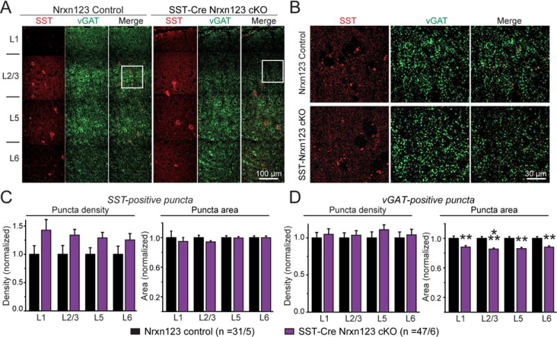
(A) Representative images (assembled from multiple overlapping views) of mPFC sections that were immunostained for SST (red) and the vesicular GABA transporter (vGAT, green). Sections were from littermate control Nrxn123 cKO (left) and SST-Cre Nrxn123 cKO mice (right). Cortical layers L1 to L6 are indicated on the left.
(B) Expansion of the boxed sections outlined in A.
(C, D) Summary graphs of the density (left) and apparent area (right) of SST+ (C) and vGAT+ synaptic puncta (D) quantified separately in each cortical layer.
Data in summary graphs are means ± SEM; statistical comparisons were performed with Student’s t-test (*P<0.05; **P<0.01; ***P<0.001; non-significant comparisons are not labeled). Numbers indicate the number of mPFC sections and mice examined. For additional data, see Fig. S7.