
Figure 19.
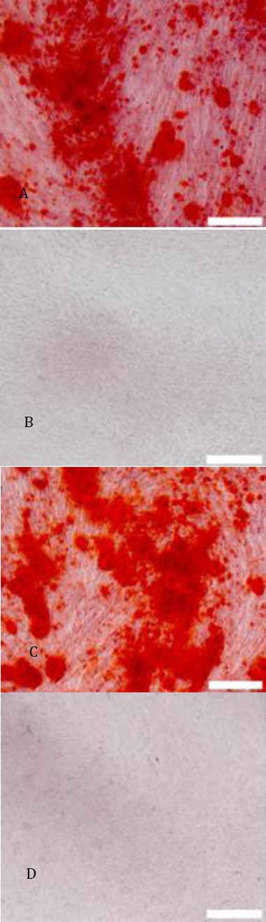
A C:hHGF-HFMSCs group and GFP-HFMSCs guoup; alizarin red staining revealed red cells at the deposition sites of calcium salt after 4 weeks of induction(40×); B D: Cells cultured for 4 weeks in basic medium instead of osteogenic differentiation media were used as the negative control. Alizarin red staining revealed that cells did not change color (40×)