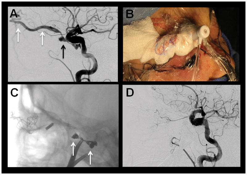
Figure 1.

(A) Pre-treatment DSA, lateral magnified view, right common carotid artery injection, showing early, rapid opacification of the right cavernous sinus (black arrow) and right superior ophthalmic vein (white arrows) consistent with a direct carotid-cavernous fistula. There is also fusiform dilatation of the cavernous right ICA representing dysplasia with an aneurysm. (B) Photograph showing the direct access provided to the right superior ophthalmic vein for coil embolization (C) Unsubtracted lateral magnified view angiography after coil embolization of the carotid-cavernous fistula showing a coil basket (white arrows) within the right cavernous sinus. (D) DSA, lateral magnified view, right common carotid injection, after coil embolization showing complete closure of the carotid-cavernous fistula without evidence of residual arteriovenous shunting.
