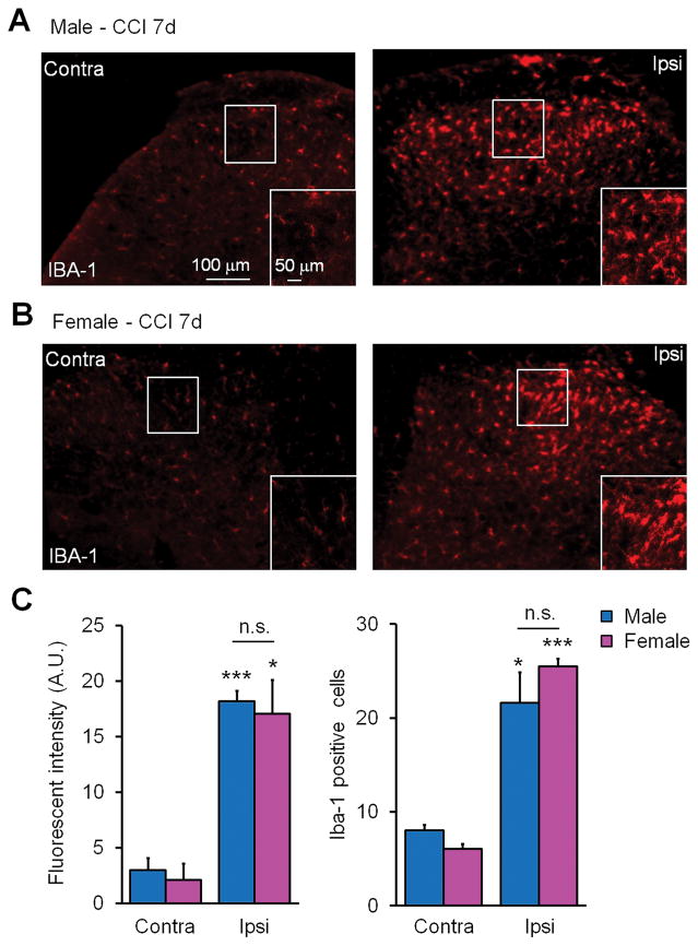
Figure 4. Spinal dorsal horn microglia display morphological activation, increased IBA-1 staining intensity, and proliferation in response to CCI in both sexes.
(A,B) Immunofluorescent staining with the microglial marker IBA-1 (red) in lumbar dorsal horn sections showing both contralateral (Contra) and ipsilateral (Ipsi) sides of male (A) and female (B) mice 7 days post-CCI. Scales, 50 and 100 μm. (C) Quantification IBA-1 immunofluorescence from mouse lumbar sections shows a significant increase in fluorescent intensity and the number of IBA-1 positive cells in the ipsilateral dorsal horn in both male and female mice. *P<0.05, ***P< 0.001, comparing ipsilateral and contralateral dorsal horn of same sex. Two-tailed Student t-test, n = 3 mice per sex per group, 3 sections per mouse.

