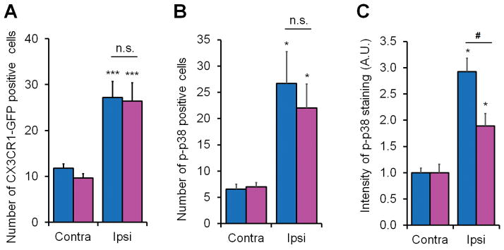
Figure 7. Nerve injury induces spinal microglial proliferation in both sexes and spinal microglial p38 activation primarily in male mice 7 days post CCI.
(A) Quantification from lumbar spinal cord sections of CX3CR1-GFP mice shows a significant increase in the number of GFP positive microglia in the medial superficial dorsal horn ipsilateral to CCI in both male and female mice. ***P< 0.001 comparing ipsilateral and contralateral of same sex, two-tailed Student t-test, n = 3 mice per sex per group. n.s., not significant. (B) Quantification of p-p38 positive cells in the medial superficial dorsal horn from lumbar spinal cord sections shows a significant increase in the number of p-p38 positive cells in in both male and female mice. *P<0.05, comparison between ipsilateral and contralateral of same sex, n = 4 mice per sex per group. n.s., not significant. (C) Quantification of p-p38 immunofluorescence in the medial superficial dorsal horn from lumbar spinal cord sections shows a significant increase in intensity of p-p38 immunofluorescence in both male and female mice and further increase in male mice. *P<0.05, comparison between ipsilateral and contralateral of same sex; #P<0.05, male vs. female, two-tailed Student t-test, n = 4 mice per sex per group. Four-five spinal cord sections were included per mouse for quantification.

