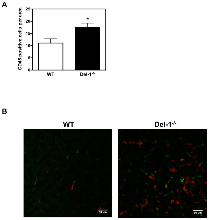
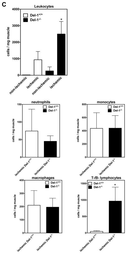
Figure 3. Del-1 deficiency enhances leukocyte infiltration of ischemic tissues.
(A) Infiltration of ischemic retinas on postnatal day 15 with CD45+ leukocytes in WT and Del-1−/− mice in the ROP model is shown. Quantification of CD45+ leukocytes in ischemic retinas by fluorescence microscopy in WT and Del-1–deficient mice (*P<0.05 vs. WT, n=4). (B) Infiltration of ischemic muscles with CD45+ leukocytes (green fluorescence) in WT and Del-1−/− mice 2 weeks after the induction of hind limb ischemia, as assessed by confocal microscopy (40X objective). The red fluorescence indicates PECAM-1+ vessels. (C) Flow cytometry of digested muscles 4 days after induction of hind limb ischemia is shown. The infiltrated leukocytes were analysed by flow cytometry for leukocyte subset markets. Data are presented as infiltrated cells/mg tissue ± SEM. (*P<0.05 vs ischemic Del-1+/+, n=5–7/group).


