Figure 6.
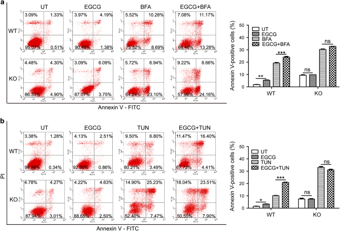
EGCG enhanced ER stress-induced apoptosis by targeting PARP16. Flow cytometry analysis with Annexin V-PI staining was performed to evaluate the percentage of apoptotic cells in EGCG combination with BFA (a) or TUN (b) or BFA/TUN treatment alone induced QGY-7703 WT and PARP16-deficient cells for 24 h. EGCG treatment significantly increased the percentage of apoptotic cells in the QGY-7703 WT cells when compared with that of BFA or TUN treatment alone. While EGCG played little or no role in the percentage of apoptotic cells in the PARP16-deficient cells compared with that of controls. Histograms showing analysis on cell apoptosis results were displayed on the right and data were shown as means±S.D. for the independent experiments. *P<0.05, **P<0.01, ***P<0.001 and NS indicated there was not statistically significant (P>0.05). BFA: 5 μg/ml; TUN: 5 μg/ml; EGCG: 100 μM.

