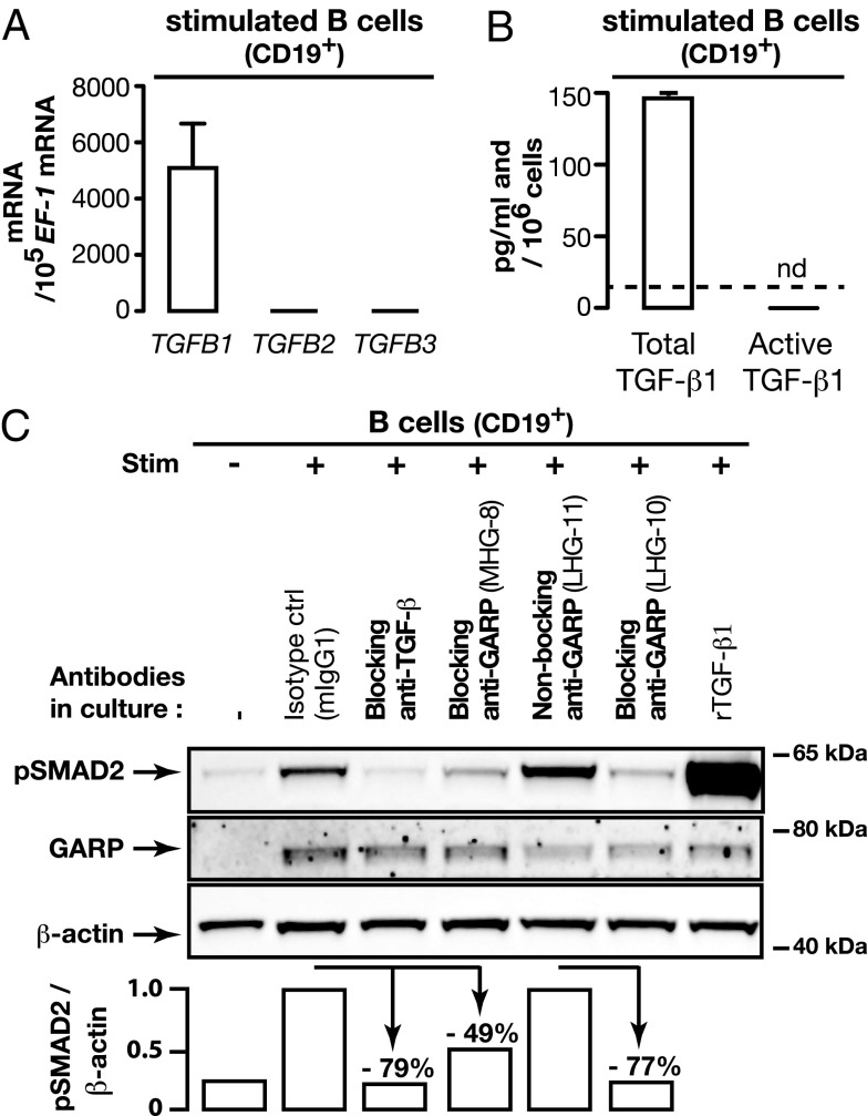
FIGURE 2.
Human B cells produce active TGF-β1 in a GARP-dependent manner. (A) Levels of TGFB1, 2 and 3 mRNA were measured in CD19+ B cells stimulated for 24 h with the mixture used in Fig. 1B. Bar graphs indicate mRNA copy numbers normalized to 105 EF-1 mRNA copies (means of duplicates + SD) as determined by quantitative RT-PCR. (B) Total and active TGF-β1 were measured by ELISA in the supernatants of B cells stimulated with the mixture used in Fig. 1B during 4 d in serum-free medium. Means of duplicates + SD. Detection limit: 16 pg/ml. nd, not detected. (C) CD19+ B cells were stimulated as in (A). The indicated Abs (10 μg/ml) were added on day 0 and 3 of the cultures, and rTGF-β1 (0.1 ng/ml) on day 3. Cells collected on day 4 were analyzed by Western blot, and luminescence signals were quantified using Bio1D software. Reduction in pSMAD2/β-actin ratios in cells treated with anti-TGF-β or the blocking anti-GARP MHG-8 Ab (both mIgG1s) are shown relative to the cells stimulated in the presence of a mIgG1 isotype control. Reduction in cells treated with the blocking anti-GARP LHG-10 Ab is shown relative to cells treated with the nonblocking anti-GARP LHG-11 (both hIgG1s). Data are representative of three independent experiments.

