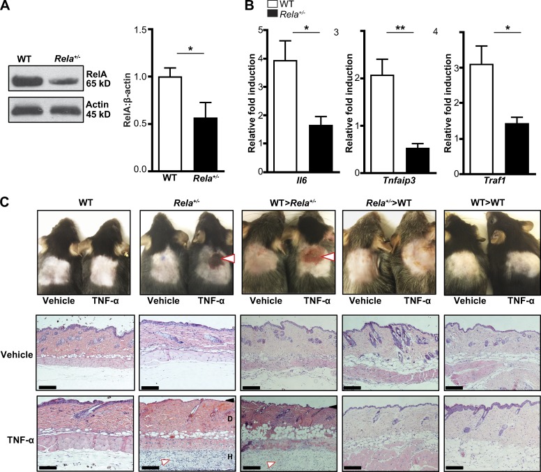
Figure 4.
RelA haploinsufficient mice develop cutaneous ulceration after TNF exposure. (A) Immunoblotting for RelA in WT and Rela+/− mice (n = 8 mice per genotype, pooled from three independent experiments). (B) qPCR analysis of NF-κB–regulated genes in WT and Rela+/− splenocytes after 24 h of TNF treatment. Gene expression was normalized to β-glucuronidase and expressed as the relative fold increase compared with unstimulated cells. Data are pooled from three independent experiments (n = 6 per genotype). (C, top) Photographs of shaved skin 24 h after s.c. injection of saline (vehicle) or TNF in the indicated genotypes. White arrows note ulceration. (Bottom) Hematoxylin and eosin stain of TNF-treated skin from WT and Rela+/− mice. The black arrow indicates loss of the epidermis. The white arrow indicates the inflammatory infiltrate. D, dermis; H, hypodermis. Bars, 500 µm. Data are from one representative experiment of three independently performed experiments. Means ± SEM are shown. *, P < 0.05; **, P < 0.01; Student’s t test.

