Figure 2.
Induction of WUS Expression by Cytokinin Treatment.
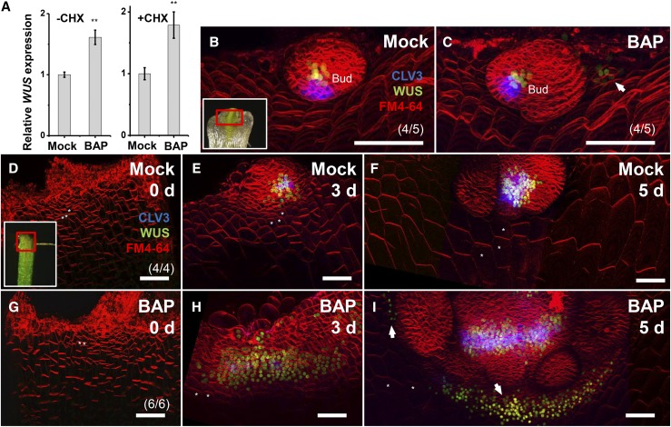
(A) A 4-h 0.89 μM BAP treatment induced WUS expression in leaf-removed shoot apex tissues in both the absence and the presence of CHX. Error bars indicate the sd of three biological replicates, run in triplicate. **P < 0.01 (Student’s t test).
(B) to (I) Time-lapse images of WUS expression in leaf axils. BAP treatment caused rapid induction of ectopic ProWUS:DsRed-N7 in mature leaf axils (C) but delayed induction in immature leaf axils ([G] to [I]).
(B) and (C) A 24-h 0.89 μM BAP treatment (C), but not mock treatment (B), induced ectopic expression of ProWUS:DsRed-N7 (green) in the leaf axil of an isolated P15 stage leaf (arrow).
(D) to (I) BAP treatment alters the WUS expression level, but not its timing. Time-lapse images showing expression of ProWUS:DsRed-N7 (green) and ProCLV3:GFP-ER (blue) in isolated P9 leaf axil centers after mock treatment ([D] to [F]) or 0.89 μM BAP treatment ([G] to [I]). BAP treatment caused the leaf axil center WUS expression domain to enlarge and activated ectopic WUS expression centers (arrows), but did not activate precocious WUS expression.
The regions bordered by the red boxes in the insets in (B) and (D) roughly correspond to the imaged region. Asterisks in (D) to (F) and (G) to (I) label the same cells in corresponding time points. The cell membrane was labeled using FM4-64 (red). Bars = 50 μm.

