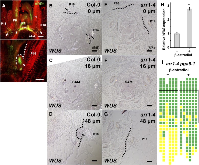
Figure 4.
Activation of WUS Expression by ARR1 Is Required for AM Initiation.
(A) Expression of ProARR1:GFP-N7 in the leaf axil. Images show longitudinal sections through a vegetative shoot apex demonstrating expression of ProARR1:GFP-N7 (green) in the leaf axil prior to (upper panel) and during (lower panel) AM initiation. Arrows indicate GFP in leaf axils. The dotted line indicates the outline of the bulged meristem. PI, propidium iodide.
(B) to (G) In situ hybridization of WUS. Images show serial transverse sections of vegetative shoot apexes in Col-0 ([B] to [D]) and the arr1-4 mutant ([E] to [G]). Arrows indicate WUS signal in leaf axils, and dotted lines indicate the outlines of leaf axils. Sections are ordered from apical ([B] and [E]) to basal ([D] and [G]). The approximate distance from the summit of the SAM to section is given in the upper right-hand corner of each image. Note that (D) and (G) are from regions more distant from the center of the same plants shown in (B) and (C), and (E) and (F). Bars in (A) to (G) = 50 μm.
(H) Expression of WUS after 8-h mock treatment or induction of ARR1ΔDDK in leaf-removed shoot tissues. Error bars indicate the sd of three biological replicates, run in triplicate. **P < 0.01 (Student’s t test).
(I) Schematic diagram of axillary buds of arr1-4 pga6-1 mutants with or without β-estradiol induction to activate WUS overexpression. See the legend to Figure 1C for a description of symbols. Green indicates the presence of an axillary bud, and yellow indicates the absence of an axillary bud. Plants were grown under short-day conditions for 15 d without treatment; leaf axil regions were treated with 10 μM β-estradiol every other day for another 15 d and then shifted to long-day conditions without treatment until axillary buds were counted. The vertical line indicates leaves initiated during β-estradiol treatment.

