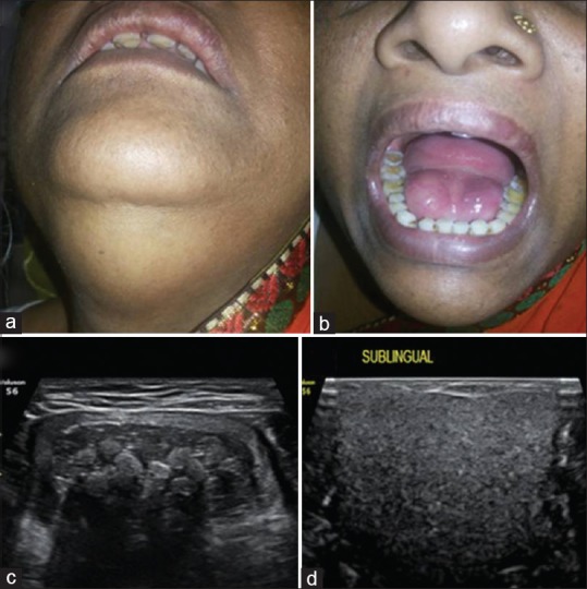
Figure 1.

Submental swelling with “double chin” appearance (a), sublingual swelling (b). Ultrasonography of submental swelling shows thick walled unilocular cystic swelling with fluid content and floating nonshadowing echogenic nodules like “stack of marbles” (c) and sublingual swelling shows unilocular cystic lesion with internal homogeneous particulate component (d)
