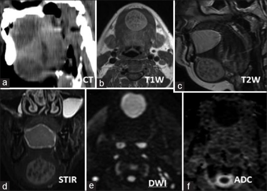
Figure 2.

Hypodense cystic sublingual and submental swelling (a) which on magnetic resonance imaging images shows T1-weighted hyperintense (b), T2-weighted/short tau inversion recovery hyperintense signal (c and d), diffusion restriction in diffusion weighted image/apparent diffusion coefficient (e and f). The submental lesion shows T1-weighted/T2-weighted/short tau inversion recovery hypointense nodules within the fluid content
