Figure 4.

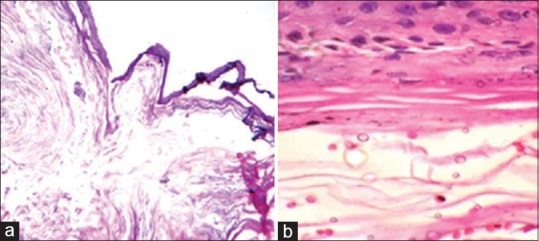
Microscopic examination of the lesions showing stratified squamous epithelial lining with accumulated keratin and underlying fibrous tissue with small blood vessels in low and high magnification (a and b)

Microscopic examination of the lesions showing stratified squamous epithelial lining with accumulated keratin and underlying fibrous tissue with small blood vessels in low and high magnification (a and b)