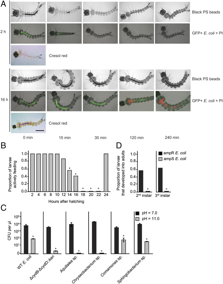
Fig. 3.
Pre- and postcritical size larvae exhibit differences in food excretion and viability of gut bacteria. (A) Images show 2-h posthatching (precritical size) or 16-h posthatching (postcritical size) first instars that were monitored over a 240-min observation period. The head of each larva is oriented to the left (anterior). (Upper) Black polystyrene (PS) beads in the midgut (Mg) and hindgut (Hg) over the 240-min observation period. The Mg and Hg are filled with beads at 0 min in 2-h and 16-h larvae. All beads have been excreted by 30 min in 2-h larvae, whereas most beads remain present after 240 min in 16-h larvae. (Middle) E. coli expressing GFP+ and PI staining of bacteria. The Mg is filled with GFP+ bacteria (green) at 0 min in 2-h and 16-h larvae. No PI staining (red) is visible, indicating bacteria are viable. All bacteria have been excreted by 30 min in 2-h larvae, whereas bacteria remain present after 240 min in 16-h larvae. Bacteria in the anterior Mg are stained by PI at 120 min, whereas bacteria in both the anterior and posterior Mg are stained at 240 min. (Lower) Two-hour and 16-h larvae at 0 min after ingestion of the pH indicator cresol red. The magenta color in the anterior Mg extending to the posterior Mg indicates a strongly alkaline pH (>10). (Scale bar, 200 μm.) (B) Proportion of larvae from 2 to 24 h posthatching that excreted all PS beads and bacteria within 30 min. An asterisk (*) indicates time points when the proportion of larvae that have excreted beads in 30 min was significantly lower than the 2-h time point (P < 0.0001, Fisher’s exact test). At least 29 larvae were assayed per time point. (C) Sensitivity of WT E. coli, ΔcydB-ΔcydD::kan E. coli, and four abundant bacterial species present in conventionally reared larvae (Aquitalea sp., Chryseobacterium sp., Comamonas sp., and Sphingobacterium sp.) to culture at pH 7 vs. pH 11. Bacteria were incubated for 2 h in neutral or alkaline LB K medium and then plated on neutral LB agar to determine the number of colony-forming units per microliter. For each species, an asterisk (*) indicates a significant difference between treatments (t test, P < 0.01). Three replicate cultures were tested per species and pH treatment. (D) Proportion of larvae that molted and successfully developed into adults when inoculated with ampicillin-susceptible (ampS) or ampicillin-resistant (ampR) WT E. coli and subsequently treated with ampicillin. Larvae were treated with the antibiotic immediately after molting to the second or third instar. An asterisk (*) indicates a significant difference between treatments (P < 0.0001, Fisher’s exact test). At least 60 larvae were assayed per instar and treatment.

