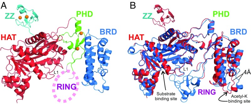
Fig. 1.
Crystal structure of the CBP catalytic core. (A) Cartoon representation of the structure of the BCHZ∆AL construct. The BRD, PHD, HAT, and ZZ domains are shown in blue, green, red, and cyan, respectively. Zinc atoms are shown as orange spheres. Electron density for the RING domain was largely absent, and its approximate location is indicated schematically by the pink oval. (B) Superposition of the CBP (red) and p300 (PDB ID code 4BHW, blue) structures showing that the acetyl-lysine binding site of the BRD is ∼4 Å closer to the HAT domain in CBP compared with p300. The acetyl-lysine binding site of the BRD and the substrate binding site of the HAT domain are indicated by arrows.

