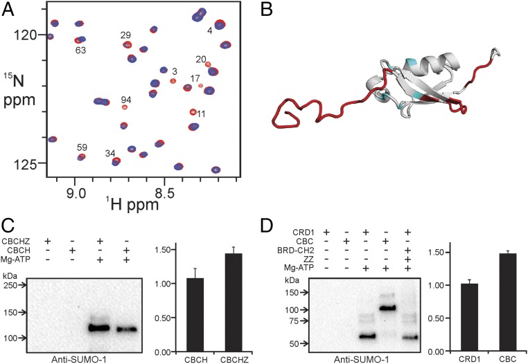
Fig. 4.
The CBP core interacts with SUMO-1. (A) Overlay of a region of the 1H–15N HSQC spectra of free 15N-labeled SUMO-1 (red) and in the presence of equimolar BCHZ∆R∆AL (blue). Cross peaks that are broadened or shifted are marked with residue numbers. (B) Mapping of the binding site (red) of the CBP core on the SUMO-1 structure (PDB ID code 1A5R). The binding site for the isolated ZZ domain, identified by Diehl et al. (25), is shown in blue. (C) In vitro SUMOylation of CRD1 repressor domain in CBCHZ and CBCH constructs. SUMOylated protein was detected using anti–SUMO-1 antibody (Left). Densitometric quantification (Right) was based on the average of five independent repeats and SEs are shown. (D) In vitro SUMOylation of GST–CRD1 and GST–CRD1–BRD–CH2. SUMOylated protein was detected by anti–SUMO-1 antibody (Left). Densitometric quantification (Right) was based on the average of five repeats, and SEs are shown.

