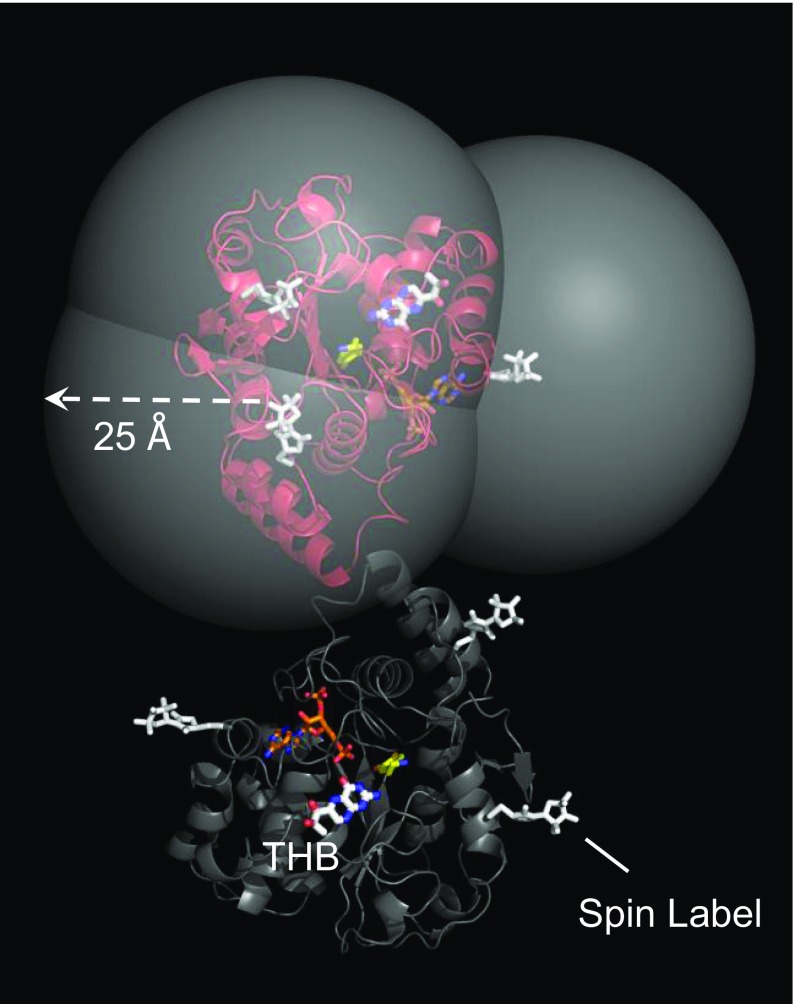
Fig. 2.
The SULT1A3 spin-label constructs. The subunits of SULT1A3 dimer are in red and gray. THB is labeled and is positioned at the catechin-binding site on the basis of screening studies (Results and Discussion). The spin labels (white) are positioned such that their paramagnetic fields can perturb the solution NMR spectra of allosteres that bind the catechin site without affecting the enzyme’s initial-rate and catechin-inhibition parameters. The carbon atoms of dopamine and PAP are yellow and orange, respectively. The semitransparent spheres center on the nitroxyl-oxygen of the spin labels, and their radii are set to the approximate maximum distance over which ligand/spin label interactions can be detected (i.e., ∼25 Å). Unlike the figure, each experimental construct has spin label attached at a single position. The design allows allostere protons to be positioned by triangulation from three spin labels.

