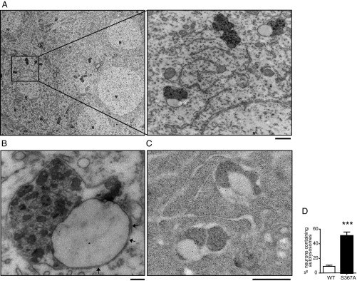
Fig. 1.
Incompletely fused autophagosomes–lysosomes are increased in brains of PS1-S367A mice. (A) Representative electron micrographs of autolysosomes in a pyramidal neuron from the CA1 area of the hippocampus from S367A knock-in mice. (Scale bar, 500 nm.) (B) Higher magnification of an unfused autolysosome. Arrows point to the double membrane. (Scale bar, 100 nm.) (C) Autolysosomes in the CA1 area of the hippocampus in PS1-S367A mice contain Cathepsin D, as revealed by immunoelectron microscopy. (Scale bar, 500 nm.) (D) Quantification of the percentage of neurons bearing autolysosomes, as seen by electron microscopy. Sixty fields from three different WT and PS1-S367A mice were used for quantification. N, nucleus. Data represent mean ± SEM. ***P < 0.001, t test.

