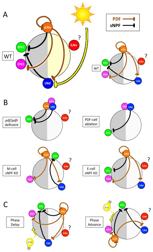
Figure 8. Model of PDF-, sNPF- and light-mediated interactions that in concert set sequential Ca2+ activity phases of the different pacemaker groups.
(A) The position of each pacemaker group on the circle indicates its Ca2+ peak phase. Both PDF and sNPF signals suppress the receivers (LNd and DN3 for PDF; DN1 for sNPF) from being active when senders (s-LNv for PDF; s-LNv and LNd for sNPF) are active. Light cycles act together with PDF to delay LNd Ca2+ phases (Figure 1A&C). (B) Loss of neuropeptide-mediated interactions caused alterations in network Ca2+ activity patterns: pdf/pdfr deficient (Figure 1D and Figure 2B), M-cell sNPF knockdown (Figure 7B), E-cell sNPF knockdown (Figure S6F), and PDF cell ablation (Figure S7). (C) Ca2+ phase shifts occurring within 24 h following 15 min light pulses suggest that light also regulates PDF and sNPF signals (Figure 5).

