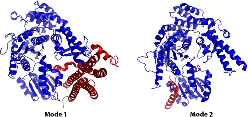
Figure 1. Binding interactions between SM and syntaxin proteins.

Mode 1 (left): The closed conformation of the mammalian neuronal syntaxin1a (red) binds in the cleft of the SM protein Munc18a (blue; Burkhardt; PDB ID 3c98). Mode 2 (right): The N-terminal peptide of yeast Sed5p binds in a hydrophobic pocket on the surface of the SM protein Sly1p (Bracher; PDB ID 1mqs). Similar N-peptide binding is observed in the Munc18c-syntaxin4 structure (Hu; PDB ID 2pjx) and in this re-refined Munc18a-syntaxin1a (not shown).
