
Figure 1. Histologic features and immunophenotype of SPCRP.
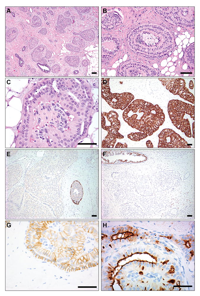
(A) Circumscribed, solid papillary tumor cell nests infiltrating around and between normal ducts. (B) Solid papillary nests with fibrovascular cores containing aggregates of foamy histiocytes. (C) Columnar cells with abundant eosinophilic cytoplasm, mild nuclear atypia, and striking reverse nuclear polarity. (D) Neoplastic epithelium demonstrating diffuse and strong expression of high molecular weight cytokeratin CK5/6. (E) Absence of myoepithelial cells around tumor nests (left) on p63 immunostain with positive internal control (right). (F) Absence of ER staining with positive internal control (upper left). (G) Absence of apical and basal E-cadherin staining with preservation of lateral membrane expression. (H) Strong expression of MUC1 in apical membranes. Scale bars, 150μm.