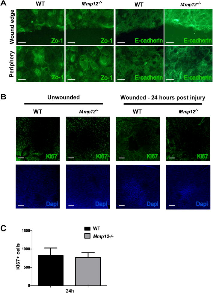
Fig. 4. MMP12 does not affect cell adhesion or cell proliferation following epithelial injury.

(A) Immunofluorescence of cell adhesion markers Zo-1 and E-cadherin in wounded primary epithelial cell cultures from WT and Mmp12-/- corneas. Images are of cells at the wound edge or peripheral to the wound edge. Scale bars: 50 μm. (B) Micrographs of representative whole-mount preparations of central corneas stained for the proliferation marker Ki-67 in unwounded and wounded (24 hours after injury) corneas. Scale bars: 100 μm. Dapi staining of cell nuclei was used to assess wound closure. (C) Quantification of Ki-67 cell counts in the central corneas of wounded WT and Mmp12-/- mice 24 hours after injury. Mean ± s.e.m. Ki-67 + cell counts are shown and are 823 (WT, n=8) and 770 (Mmp12-/-; n= 16).
