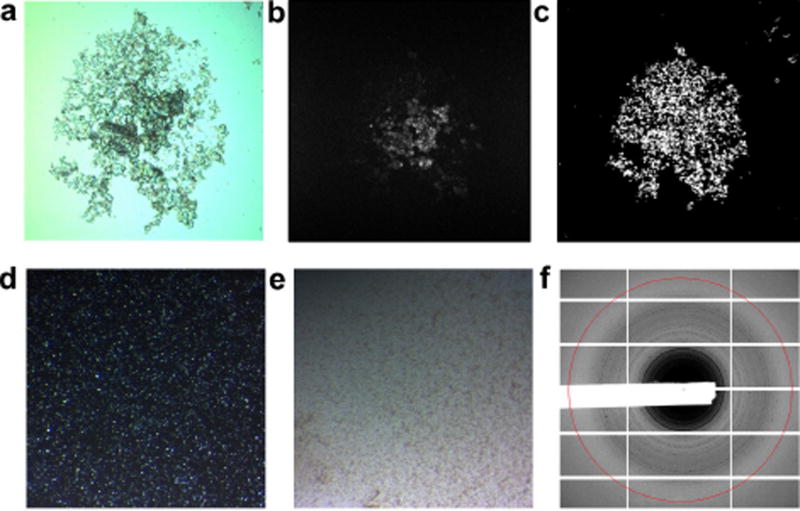
Extended Data Figure 1. Characterization of RNA crystals.
Microcrystals of apo-rA71 were grown using batch crystallization as described in Methods. A SONICC Imager (Formulatrix) was used to image each sample (0.5–1.0 µl) of crystals using three different methods: visible light (a); UV-TPEF (b); and second-order nonlinear imaging of chiral crystals (SONICC) (c). d, Crystal samples were observed using a stereomicroscope (Zeiss) under cross-polarized light. e, Without cross-polarization, crystals were barely observable. f, The relative quality of the crystalline samples was measured by powder X-ray diffraction (APS beamline 19-ID), with a maximum observable resolution of approximately 6 Å. The resolution ring (red) corresponds to 6.8 Å.

