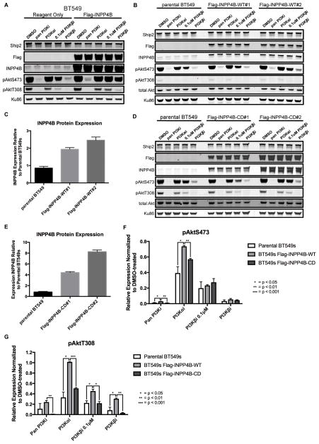
Figure 4. Effect of wild type or catalytically-dead INPP4B overexpression on pAkt levels in BT549 cells.
A, BT549 cells transfected with a plasmid encoding Flag-INPP4B or reagent only, and 48 hours later treated for 1 hour with either DMSO or 1μM inhibitor except where it is noted that PI3K-βi was used at 0.1μM. Ku86 was used as a loading control. B, parental BT549, BT549 Flag-INPP4B-WT#1 or BT549 Flag-INPP4B-WT#2 cells were treated for 1 hour with either DMSO or 1μM inhibitor except where it is noted that PI3K-βi was used at 0.1μM. C, average quantified INPP4B protein expression, normalized to Ku86 loading control and then to the DMSO-treated parental BT549 cells. D, parental BT549, BT549 Flag-INPP4B-CD#1 or BT549 Flag-INPP4B-CD#2 cells were treated for 1 hour with either DMSO or 1μM inhibitor except where it is noted that PI3K-βi was used at 0.1μM. E, average quantified INPP4B protein expression, normalized to Ku86 loading control and then to DMSO-treated parental BT549 cells. F–G, quantified pAktS473 (F) and pAktT308 (G) in BT549 parental cells, Flag-INPP4B-WT cells (Flag-INPP4B-WT#1 and -WT#2 pooled) and BT549 Flag-INPP4B-CD cells (Flag-INPP4B-CD#1 and -CD#2 pooled), normalized to the DMSO-treated sample for each cell line. Cells were treated for 1 hour with either DMSO or 1μM inhibitor except where it is noted that PI3K-βi was used at 0.1μM. (pan PI3Ki: GDC-0941, PI3K-αi: BYL719, PI3K-βi: AZD6482)

