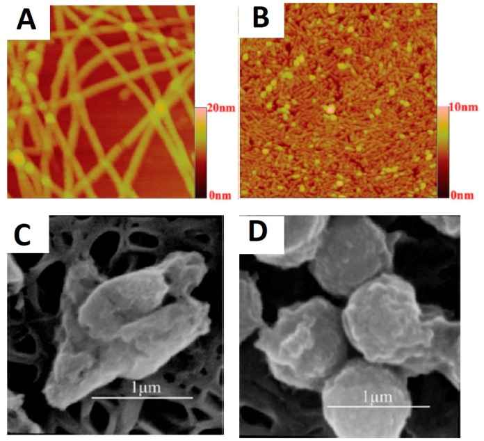
Figure 4.
Nanostructures formed by peptide self-assembly as revealed by topographical AFM micrographs formed by: (A) A6K; and (B) A9K. Scanning electron micrographs of: (C) E. coli; and (D) S. aureus treated by A9K. Figures reproduced from [88] with permission from ACS.

