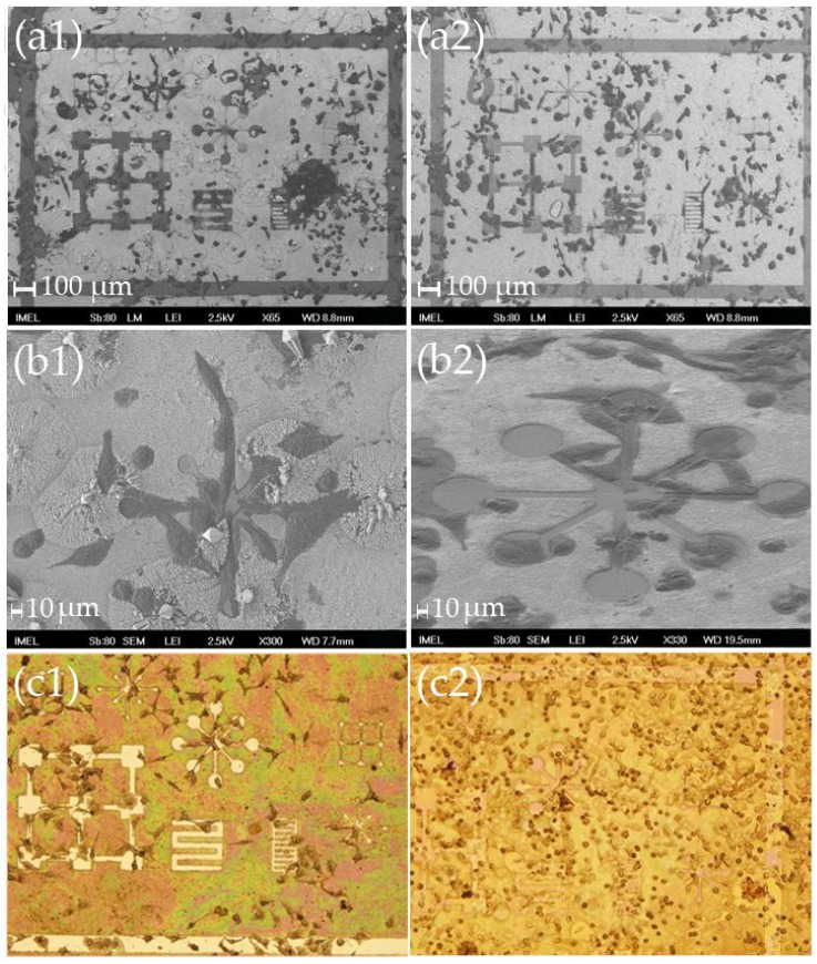
Figure 3.
(a); (b) SEM images at two different magnifications (scale bars: 100 μm and 10 μm, respectively) and (c) optical microscope images of HeLa cells after 2 days (left column), and 4 days (right column) in culture on modified nitride samples “S” with small free patterns, where it can be seen that despite the limited flat surface the cells tend to avoid the nanostructured areas and try to “squeeze” inside the patterns. On day 4, the cells that adhered onto the nanostructures were no longer elongated and obtained a more spherical shape. (a1); (b1); and (c1) were obtained after 2 days in culture, while (a2); (b2); and (c2) were obtained after 4 days in culture.

