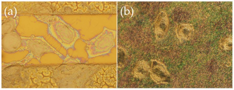
Figure 5.
High magnification (×100) optical microscope images in dark field of HeLa cells after 4 days in culture on modified nitride samples that have adhered (a) on the nitride surface; and (b) onto the nanostructures. In (a), one can see the extensive filopodia network, while in (b) the more spherical shape of the cells can be discerned. The colorful specs are the tips of the ZnO nanostructures as seen in dark field.

