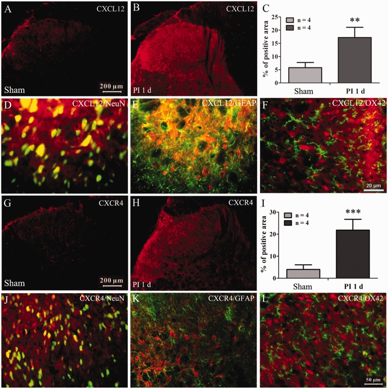
Figure 2.
Distributions and cell-types of CXCL12 and CXCR4 expressed in spinal dorsal horn. (a) to (c) The immunofluorescence staining pictures showing increased expression of CXCL12 in spinal dorsal horn. **P < 0.01 versus sham group, Student's t-test. (d) to (f) Representative pictures showing the CXCL12 colocalized with neuronal marker NeuN (d) and astrocytic marker GFAP (e), but not microglia marker OX42 (f). (g) to (i) The immunofluorescence staining pictures showing an increased expression of CXCR4 in spinal dorsal horn. ***P < 0.01 versus sham group, Student's t-test. (j) to (l) Representative pictures showing the CXCR4 exclusively colocalized with neuronal marker NeuN (j), but not astrocytic (k) and microglia marker (l).
PI: Plantar incision.

