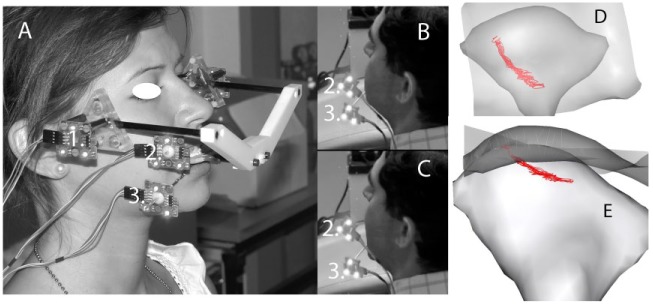
Figure 1.
Jaw-tracking data collection for dynamic stereometry where (A) shows a subject wearing the custom oral appliance connected to the reference system with light-emitting diodes (LED) (1). LED are also connected to the maxillary (2) and mandibular (3) teeth via custom splints affixed temporarily to the teeth. A subject’s jaw position at (B) maximum intercuspation and (C) maximum opening is shown during jaw movement as this is tracked and recorded by cameras located outside the field of view to the subject’s left. Dynamic stereometry results in (D) superior and (E) frontal views after reconstruction of temporomandibular joint (TMJ) anatomy and position data are combined and centroid of the stress field identified at each position. Right TMJ condyle and semi-transparent image of fossa and eminence are shown, where the paths of the centroid of the stress field between maximum intercuspal position and maximum jaw opening can be seen in red.

