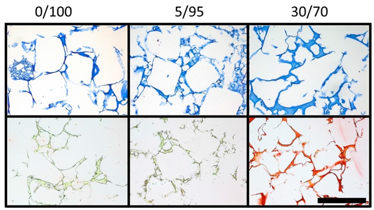
Figure A1.
DMMB and picrosirius red staining of empty scaffolds of different composition (columns): DMMB staining for glycosaminoglycans (e.g., HA) at ×10 magnification (upper row) and picrosirius red staining for collagen (e.g., gelatin) content (scale bar = 500 µm). Scaffolds with higher gelatin content are more intensely stained by picrosirius red, while homogenous distribution of the components within the scaffolds is visualized.

