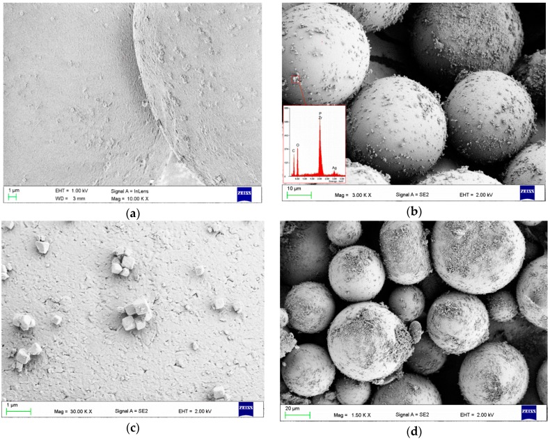
Figure 2.
Representative SEM images presenting the surface of the PMMA spheres after filler introduction: (a) sphere surface after milling with 0.25% filler; (b) surfaces of spheres with 2% filler and the corresponding energy-dispersive X-ray spectroscopy (EDS) spectrum, which confirmed the presence of zirconium, phosphorus and silver; (c) smaller aggregates of filler particles; (d) spheres covered to a large extent by filler particles when 4% filler was introduced.

