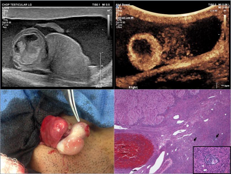
Figure 1.

15-year-old phenotypic male who presented with testicular rupture. (A) Right testicular rupture with upper pole mass and hematocele. (B) After intravenous administration of ultrasound contrast there is marked peripheral enhancement of the right upper pole mass. The lower half of the testis does not demonstrate homogenous enhancement. (C) Intraoperative gross pathology demonstrating the abnormal round mass at the superior pole of the right testis (indicated by forceps). (D) Ovarian stroma with primary and secondary follicles (→), corpus luteum in upper left corner, and corpora albicans in the lower center, (4× magnification; Hematoxylin and eosin stain); inset: secondary follicle, (40× magnification; Hematoxylin and eosin stain)
