
Figure 1. Phenotypic analysis of position of cecum in wildtype (WT) and heterozygous (HET) Fgfr2W290R mice.
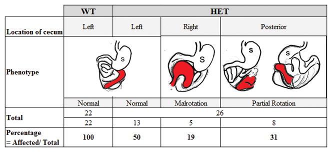
The position of the cecum in the abdominal cavity is colored in red, as viewed ventrally. In wildtype (WT) and 50% of mutant mice, the cecum lies across the abdominal cavity, stretching from left to right (denoted under “left” column). In 19% of the mutant mice, the cecum appears from right to left (“Right” column). In the remaining 31% of the mutants, the cecum is located towards the posterior aspect of the contents of the abdominal cavity (“Posterior” column where figures on left and right represent ventral and dorsal views, respectively).