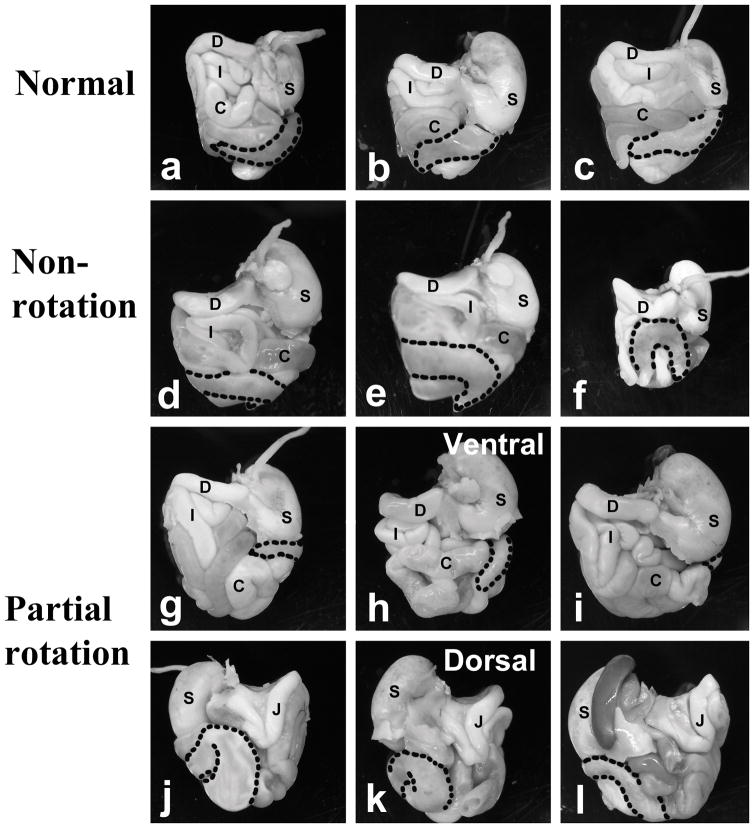
Figure 2. Position of cecum in the abdominal cavity of Fgfr2W290R mice.
Abdominal contents were removed en masse from mutant mice and photographed after removing the overlying liver (a – i: ventral views; j – l: dorsal views of same guts as shown in g–i). All wildtype (WT) mice and 50% of the Fgfr2W290R heterozygous (HET) mice presented with the cecum lying ventrally across from the left side, with the ends of the pouched-like cecum facing right (a – c are examples from three mice). In the partial rotation group (g & j, h & k, and i & l are guts from the same embryos, photographed from the ventral and dorsal views, respectively), the cecum is barely visible on the ventral surface and the bulk remains dorsally. C- colon; D- duodenum; I- ileum; J-jejunum; S-stomach; stippled line- outline of cecum.

