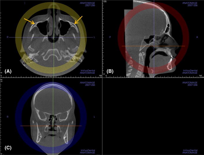
FIGURE 3.

Visualization of the ZMS: in the sagittal view, the horizontal cursor (orange line) is placed at the tip of the nose parallel to the palatal plane (B). Note that in the axial view, the zygomaticomaxillary sutures are seen bilaterally (A) (arrows). The vertical cursor (green line) should be positioned on the midsagittal plane of the patient (C) [Colour figure can be viewed at wileyonlinelibrary.com]
